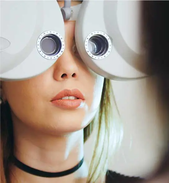
Advanced Diagnostic Technologies and Testing - We See More So That You May Never See Less.
A common misconception is that all eye exams are created equal. Unlike big box stores and many retail-focused shops, eye-deology Vision Care invests in the modern technologies required to provide state-of-the-art comprehensive eye exams and eye care. Why is this important? Because advanced technologies enable our optometrists to see more and promote the early detection of eye diseases that can cause irreversible vision loss. Like how dentists use x-rays, gynecologists use ultrasounds, and neurosurgeons use an MRI, our optometrists use technologies such as wide-angle retinal (fundus) photography, Optical Coherence Tomography (OCT), corneal topography, and digital dry eye analysis to provide the best possible care to patients.
Schedule An Exam Our Optometrists
(780) 473-6123
Eye-deology Vision Care
Wide Angle Retinal Photography Seeing More Of The Eye
Enabling our optometrists to see more helps ensure that our patients will never see less. Wide-angle retinal (fundus) photography enables our optometrists to see more - more detail and more of the eye at once. Our unique wide-angle confocal fundus camera uses a white light LED source that uses the entire visible spectrum to illuminate the retina and capture fundus images. By having retinal images, in colours close to reality, our optometrists benefit from the most accurate perception of fundus anatomy. Benefits to patients and doctors include:
- Early Detection. Early detection of macular and retinal diseases often depends on distinguishing between healthy features and potential signs of abnormalities.
- Enhanced View. Ability to scan through cataracts and other media opacities.
- Comprehensive View. The wide-field capability of up to 150° enables detection of small details and changes in both the periphery and central region.
- Change Detection. Imagery essential to properly document, compare, and monitor changes in the macula and retina.
- Reduces Dilation Need. The technology enables our doctors to work with small pupils without the need for dilation.
Wide Angle Retinal Photography »
Schedule An Eye Exam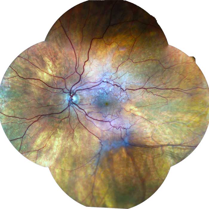
Eye-deology Vision Care
Optical Coherence Tomography: Seeing Ocular Tissues
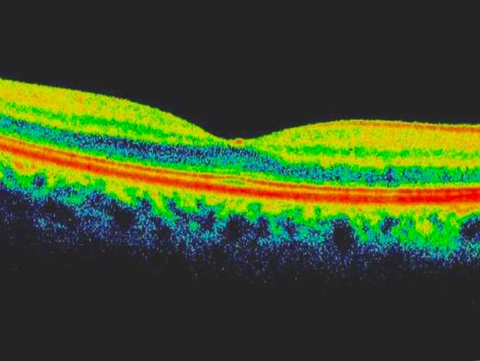
Optical Coherence Tomography (OCT) is a non-invasive diagnostic technique that renders a cross-sectional view of the retina, much like an x-ray of a tooth, except that it only uses white light. OCT utilizes a concept known as interferometry to create a cross-sectional map of the retina that is accurate to within at least 10-15 microns. OCT is useful in the diagnosis of many retinal conditions. In general, lesions in the macula are easier to image than lesions in the mid and far periphery. OCT can be particularly helpful in diagnosing:
- Macular hole
- Macular pucker/epiretinal membrane
- Vitreomacular traction
- Macular edema and exudates
- Detachments of the neurosensory retina
- Retinopathy / Age-Related Macular Degeneration
- Retinoschisis
- Pachychoroid
- Choroidal tumours
- Glaucoma
- Optic neuritis
- Non-glaucomatous optic neuropathies
- Alzheimer's disease
- Anterior segment
Eye-deology Vision Care
Corneal Topography: Mapping The Surface of The Eye
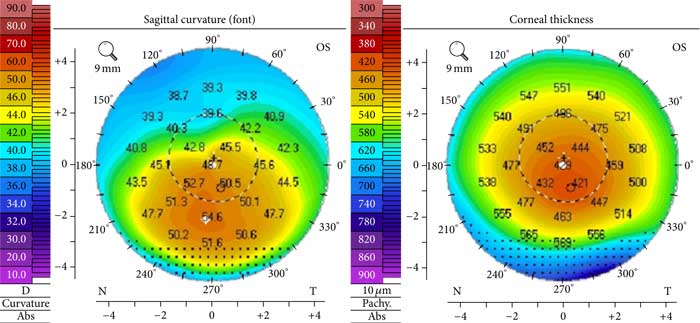
Corneal topography is a non-invasive medical imaging technique used for mapping the surface of the outer portion of the eye - the cornea. Since the cornea is responsible for approximately 70% of the eye's refractive power, its topography influences both vision quality and overall corneal health. The three-dimensional map is a valuable diagnostic tool for optometrists that is produced in seconds and is painless. Computerized corneal topography is a diagnostic tool used to:
Computerized corneal topography is performed to:
- Screen for the presence of keratoconus and analyze the degree of corneal steepness.
- Monitor corneal changes.
- Assess patients for cross-linking and mini asymmetric radial keratotomy.
- Custom fit contact lenes
Eye-deology Vision Care
Dry Eyes: Jenvis Dry Eye Report
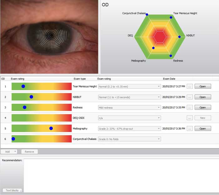
eye-deology Vision Care optometrists use a corneal topographer that enables them to examine the following to determine the cause of dry eye symptoms:
- Meibomian Glands. The dysfunction of meibomian glands is the most frequent cause of dry eye. Morphological changes in the gland tissue are made visible and can be classified.
- Non-Invasive Tear Film Break-Up Time. A non-invasive scan that measures and maps tear film breakup across the surface of the eye.
- Tear Meniscus Height. Tear meniscus height is precisely measured along the edge of the bottom lid using an integrated digital ruler and magnification options.
- Tear Film Dynamics. An observation of the tear film particle flow to assess tear film viscosity.
- Lip Layer Amount & Health. The interference colours of the lipid layer and their structure are made visible and recorded. The thickness of the lipid layer is assessed based on its structure and colour.
- Redness. Evaluation of the bulbar and limbal degree of redness. The R-Scan detects the blood vessels in the conjunctiva and evaluates the degree of redness.
Eye-deology Vision Care
Night Vision: Testing and Assessment
Night vision is the ability to see in low light conditions. Differences between daytime vision and night vision include:
- Pupil size increases to allow in more light in darker environments.
- A different, more sensitive type of cell in the eye, called rod cells, collects the light for night vision.
- Night vision is predominantly black and white (i.e., shades of gray).
- Colour vision is poor in very low-light conditions.
Police officers often need to undergo regular night vision testing. To assess night vision, our optometrists will perform a comprehensive eye exam administer a Nigh Vision test, using a chart such as the Pelli-Robson Contrast Sensitivity Chart. It is similar Snellen letter chart, but with letters in different shades of grey. This test measures how well an individual can see the contrast between white paper and light grey shapes.
Schedule A Night Vision Test
Our Edmonton Optometrists
Searching for an optometrist in Edmonton? Our experienced Edmonton eye doctors use advanced modern technologies and devote upwards of 500% more time towards providing personalized patient care than elsewhere so that they can see more and ensure that you may never see less. Position yourself to see the future with a visit to our eye clinic and Edmonton's best eye care!

Dr. Jennifer Ash, OD
Dr. Jennifer Ash is the Resident Optometrist at Eye-deology Vision Care. Dr. Ash provides patient care 5 days a week. Read more about Dr. Ash.

Dr. Ruhee Kurji, OD
Dr. Ruhee Kurji is an Associate Optometrist at Eye-deology Vision Care. Dr. Kurji provides patient care Tuesdays & Fridays. Read more about Dr. Kurji.

Dr. Jade McLachlin, OD
Dr. Jade McLachlin is an Associate Optometrist at Eye-deology Vision Care. Dr. McLachlin provides patient care 5 days a week. Read more about Dr. McLachlin.

Dr. Tania Mathews, OD
Dr. Tania Mathews is an Associate Optometrist at Eye-deology Vision Care. Dr. Mathews provides patient care 2 days a week. Read more about Dr. Mathews.
Learn Why Our Edmonton Optometrists Are The Best!


